Prostate Cancer
Prostate cancer is a malignant tumor that develops in the prostate gland in men. It is very common and has increasingly been diagnosed among younger patients in recent years [1-3]. In many cases, it is a chronic disease that affects various aspects of the patient’s life, ultimately leading to a decline in both quality of life and functioning. It can metastasize to nearby lymph nodes or distant organs, often to bones, and less frequently to the lungs, brain, and liver [4].
Diagnosis of Prostate Cancer – Current Standard
Currently, the gold standard for detecting and locating cancerous changes is MRI (magnetic resonance imaging). Additional imaging diagnostics for oncology patients include PET/CT scans. This method is considered safe, and radiopharmaceuticals such as 11C/18F-choline, 11C-acetate, 18F-fluciclovine, and 68Ga/18F-PSMA are used. These drugs are harmless to the patient, with doses tailored individually based on the patient’s body weight and are primarily excreted by the kidneys. More information on radiopharmaceuticals can be found here. Our BioSkaner facility offers both prostate MRI and PET/CT scans. Both can be performed privately or free of charge under the NFZ agreement. Detailed information on current appointments and examination prices can be found on the dedicated pages for MRI or PET/CT scans.
68Ga-PSMA-11
The radiopharmaceutical 68Ga-PSMA-11 has the highest sensitivity and specificity in the diagnosis of prostate cancer. PET/CT with PSMA has higher value than standard methods such as MRI, abdominal CT, bone scintigraphy, or PET/CT with radiolabeled choline. Unfortunately, although the radiopharmaceutical 68Ga-PSMA-11 is recommended by the European Association of Urology, due to its limited availability, it has not yet been introduced as a standard procedure [5, 6]. However, scientific studies indicate that the implementation of 68Ga-PSMA-11 PET in diagnostics results in a change in therapeutic decision in 51-76% of patients [7, 8, 9].
Importantly, the use of 68Ga-PSMA-11 significantly increases the ability to detect early recurrence, as it can detect PSA values <1 ng/ml (compared to 11C/18F-choline at PSA 1-2 ng/ml in 5-24% of patients), and the resolution in detecting lymph node metastases is very high, up to 3 mm (from 2.4 mm in diameter) [10, 11, 12].
Researchers highlight the need to replace conventional imaging methods, such as CT and bone scintigraphy, with PSMA PET/CT in high-risk patients before initiating radical treatment [13]. This has also been emphasized in studies verified by histopathological evaluation of postoperative changes [14].
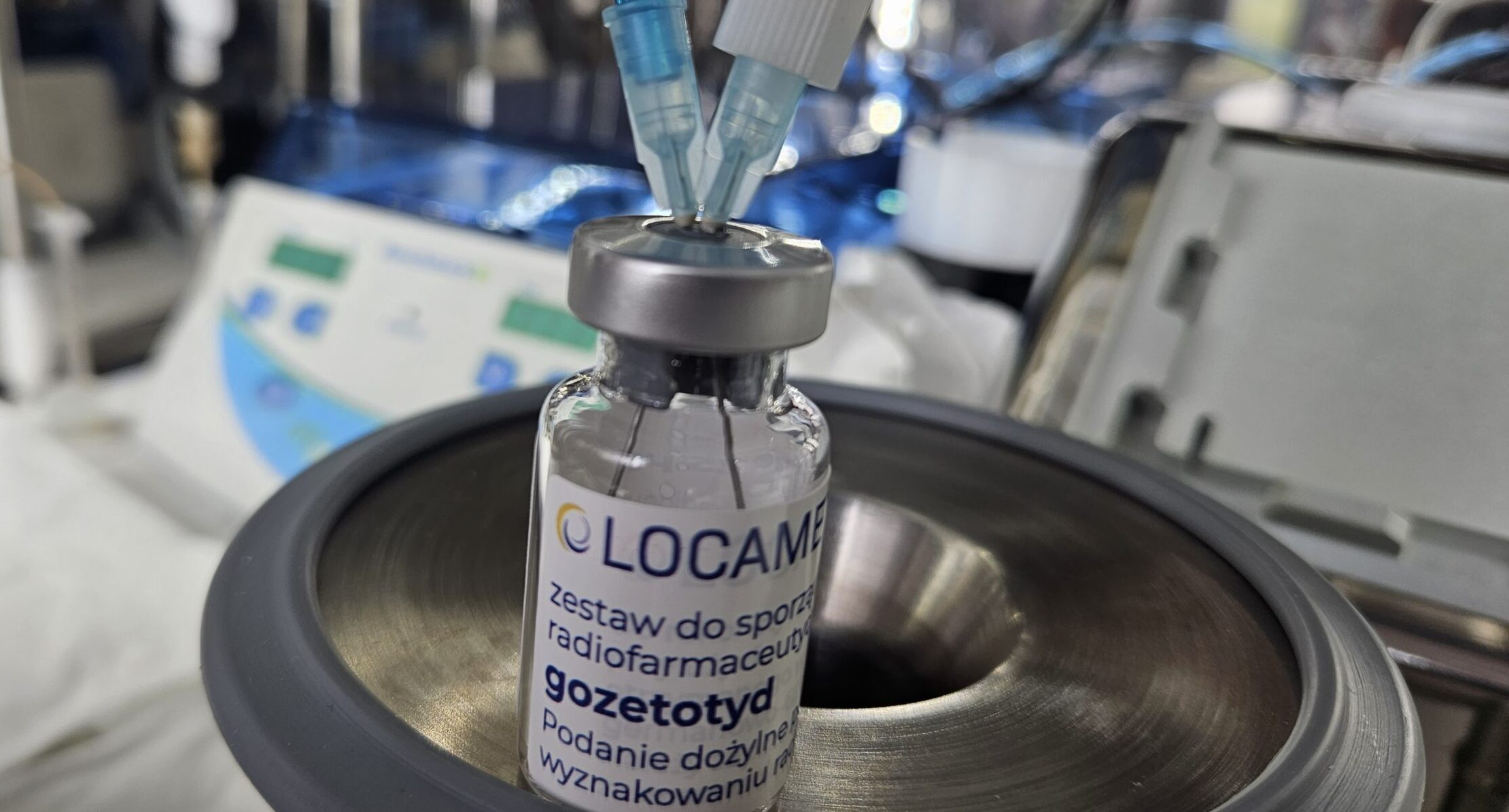
Since December 2022, we have been able to obtain the 68GaPSMA-11 radiopharmaceutical in the radiochemical laboratory of our BioSkaner center. This drug is labeled with the radioactive element 68-gallium and the PSMA-11 ligand, which binds to prostate cells. The PSMA-11 ligand serves as a carrier and enables the localization of the radiotracer at the target site in the patient’s body. It has a short half-life of 68 minutes. The drug is administered intravenously in trace amounts, delivering a minimal dose of ionizing radiation that does not affect the patient’s well-being. It is ideal for diagnostics because it is quickly excreted from the patient’s body, mainly through the kidneys [15, 16].
PET/MR with PSMA
Since 2011, isotope diagnostics using PET/MR technology have also been available worldwide. PET/MR molecular imaging provides extensive data from both PET and MR scans, and only a low dose of ionizing radiation from the applied radiotracer [17, 18]. Our BioSkaner center has been performing PET/MR scans since 2015. More information about the innovative PET/MR scan can be found here. Despite its introduction for human use over a decade ago, PET/MR technology remains a novel method.
Combining whole-body PET and MR in one examination increases the accuracy of assessing the extent of changes and the ability to detect small lesions, even at very low PSA values, such as < 0.2 ng/ml. The detection rate of recurrence is higher for PSMA PET/MR [19, 20].
Early scientific reports indicated the value of PSMA PET/MR surpassing that of PSMA PET/CT. We have discussed the differences between PET/MR and PET/CT in articles on PET/MR examinations and on the page comparing these two techniques. In a systematic review comparing 3182 publications on PET/MRI for oncological indications, PET/MRI technology was found to have the highest diagnostic value compared to PET/CT in detecting recurrence in prostate cancer patients [21].
Currently available study results indicate a very high value of PSMA PET/MR – sensitivity up to 96%, specificity up to 100% [5, 22, 23, 24]. Notably, radiotherapy as a method of radical treatment is more effective when the recurrence is localized at the lowest possible PSA values, ideally < 0.2 ng/ml [25, 26]. It should be added that our BioSkaner center meets the highest safety standards for examinations and has EARL accreditation.
The method combining PET/MRI in one examination is emphasized as the most valuable among all available techniques used in the diagnosis of patients with biochemical recurrence of prostate cancer [9]. In the case of a significant contraindication from the patient, such as severe claustrophobia, we can perform the examination under general anesthesia at the BioSkaner facility.
Unfortunately, due to its innovation, PET/MR examinations are not yet reimbursed. Therefore, examinations are carried out only for a fee. The current price list for examinations can be found here. Additionally, our two laboratories, LOM Scan and BioSkaner, have introduced a joint offer that allows for a reduction in the cost of PET/MR examinations.
Prostate cancer develops very slowly and may not show symptoms for many years. Therefore, a quick and precise assessment of the stage of cancer progression, both before starting treatment and when recurrence is suspected, is essential for making optimal therapeutic decisions [11, 27]. We invite you to take advantage of the extensive diagnostic offer of our BioSkaner facility.
Literature
- Bray F., Ferlay J et al. Global cancer statistics 2018: GLOBOCAN estimates of incidence and mortality worldwide for 36 cancers in 185 countries. CA Cancer J Clin. 2018 Nov;68(6):394-424.
- European Cancer Information System.
- Krajowy Rejestr Nowotworów.
- Wysocki W. – Rak Prostaty | Onkologia mp.pl.
- Mottet N., van den Bergh R.C.N. et al. EAU-ESTRO-ESUR-SIOG Guidelines on Prostate Cancer. Eur Urol. 2017 Apr;71(4):630-642.
- Annunziata S., Pizzuto D.A. et al. Diagnostic Performance of PET Imaging Using Different Radiopharmaceuticals in Prostate Cancer According to Published Meta-Analyses. Cancers (Basel) 2020 Aug. 4;12(8): E2153.
- Scosyrev E., Messing E. M. et al. Prostate cancer in the elderly: frequency of advanced disease at presentation and disease-specific mortality. Cancer, 2012. 118: 3062.
- Morigi, J.J., Stricker P. D. et al. Prospective Comparison of 18F-Fluoromethylcholine Ver-sus 68Ga-PSMA PET/CT in Prostate Cancer Patients Who Have Rising PSA After Cura-tive Treatment and Are Being Considered for Targeted Therapy. J Nucl Med, 2015. 56: 1185. World J Urol. 2018 Apr;36(4):519-527. doi: 10.1007/s00345-018-2182-1. Epub 2018 Jan 17.
- Corfield J., Perera M. et al. 68Ga-prostate specific membrane antigen (PSMA) positron emis-sion tomography (PET) for primary staging of high-risk prostate cancer: a systematic review. World J Urol. 2018 Apr;36(4):519-527. doi: 10.1007/s00345-018-2182-1. Epub 2018 Jan 17.
- Ceci F., Herrmann K. et al. Impact of 11C-choline PET/CT on clinical decision making in recurrent prostate cancer: results from a retrospective two-centre trial. Eur J Nucl Mol Imaging 2014 Dec;41(12):2222-31.
- Giovacchini G., Picchio M., et al. Predictive factors of 11C-choline PET/CT in patients with biochemical reccurence. Eur J Nucl Med Mol Imaging 2015.
- Afshar-Oromieh A., Avtzi E. et al. The diagnostic value of PET/CT imaging with the (68)Ga-labelled PSMA ligand HBED-CC in the diagnosis of recurrent prostate cancer. Eur J Nucl Med Mol Imaging. 2015 Feb;42(2): 197-209.
- Hofman Z, Lawrentschuk N. et al. Prostate-specific membrane antigen PET-CT in patients with high-risk prostate cancer before curative-intent surgery or radiotherapy (proPSMA): a pro-spective, randomised, multi-centre study. MBBS Lancet 2020 VOLUME 395, ISSUE 10231, P1208-1216, APRIL 11, 2020.
- Nandurkar R., van Leeuwen P. 68Ga-HBEDD PSMA-11 PET/CT staging prior to radical prostatectomy in prostate cancer patients: Diagnostic and predictive value for the biochemical response to surgery. Br J Radiol. 2019 Mar;92(1095):20180667.
- Sandgren K., Johansson L. et al. Radiation dosimetry of [68Ga]PSMA-11 in low-risk prostate cancer patients. EJNMMI Physics (2019) 6:2.
- Broski S. M., Goenka A. H. et al. Clinical PET/MRI: 2018 Update. AJR Am J Roentgenol. 2018 Aug;211(2):295-313.
- Martin O., Schaarschmidt B. et al. PET/MRI Versus PET/CT for Whole-Body Staging: Results from a Single-Center Observational Study on 1,003 Sequential Examinations Journal of Nu-clear Medicine, August 2020, Vol. 61:8, pp. 1131-1136.
- Josep M. Martí-Climent1,2*, Elena Prieto et al. Effective dose estimation for oncological and neurological PET/CT procedures EJNMMI Research (2017) 7:37.
- Hoffmann M. A., Wieler H. J., et al. The Impact of 68Ga-PSMA PET/CT and PET/MRI on the Management of Prostate Cancer. Urology. 2019 Aug;130:1-12.
- De Visschere P., Standaert C. et al. A Systematic Review on the Role of Imaging in Early Recurrent Prostate Cancer. Eur Urol Oncol 2019 Feb;2(1):47-76.
- Morsing A., Hildebrandt G. et al. Hybrid PET/MRI in major cancers: a scoping review. Re-ceived: 27 March 2019/Accepted: 13 June 2019 # Springer-Verlag GmbH Germany, part of Springer Nature 2019.
- Barbosa F., Queiroz M., et al. Clinical perspectives of PSMA PET/MRI for prostate cancer. Clinics 2018, 73, 1–9.
- Keidar Z., Gill R. et al. 68Ga-PSMA PET/CT in prostate cancer patients. Cancer Imaging 2018.
- Sachpekidis C., Kopka K. et al.. „68Ga-PSMA-11 dynamic PET/CT imaging in primary prostatę cancer” Clin Nucl Med. 2016;41:473-479.
- Pfister D., Bolla M. et al. Early salvage radiotherappy following following radical prostatectomy. Eur Urol. 2014 Jun;65(6):1034-43.
- Tendulkar R. D., Agrawal S. et al. Contemorary update of a multi-institutional Predictive Nomogram for Salvage Radiotherapy After Radical Prostatectomy J Clin Onco 2016 Volume 34, Issue 30.
- Liu D., Lehmann P. et al. Active surveillance versus surgery for low risk prostate cancer: a clinical decision analysis. J Urol, 2012. 187: 1241. 55.

